
Our Medical Services
Call 031 581 2546
Our Services
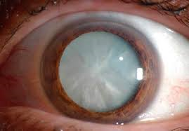
Cataract surgery
A cataract is when the natural lens in your eye becomes cloudy
Read More
It is caused by ageing, diabetes, medications such as steroids, eye injury and prolonged exposure to the sun’s UV-B light. A cataract is when the natural lens in your eye becomes cloudy.Cataracts may cause blurred or dull vision and difficulty focusing, colours that seem faded, glare, not being able to see well at night and frequent changes of prescription glasses. If the cataract makes it difficult for you to see well enough to carry out your daily activities, the cataract may need to be removed. Surgery is the only way to remove a cataract. During the operation the cloudy lens (cataract) is removed and replaced with an artificial lens implant. Cataract surgery is usually performed under light sedation and local anaesthesia. If the cataract is not removed, your vision will gradually get worse.
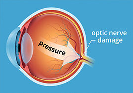
Glaucoma
Glaucoma is the leading cause of irreversible blindness worldwide
Read More
In South Africa, it affects 4 in 50 people over the age of 40 years. Glaucoma is a condition characterized by loss of vision that results from damage to the optic nerve, which usually occurs when the pressure within the eye is high, although the pressure can also be within the normal range. Glaucoma is a progressive disease without appropriate treatment and the damage to the optic nerve and vision is irreversible. It has been described as the “sneak thief of sight” because there are usually no symptoms early on in the disease. There is no cure for glaucoma however progressive vision loss and blindness is preventable if diagnosed and treated early. Regular eye examinations and glaucoma screening is therefore important in the early diagnosis and treatment of the condition. Those at risk of developing glaucoma include age over 40 (risk increases as we age), family history of glaucoma, African and Asian race group, myopia (short-sightedness) and diabetics. Diagnosis includes measurement of the intraocular pressure, assessment of the optic nerve clinically as well as performing a scan of the optic nerve and a visual field examination. Treatment options include eye drops, laser and surgery if medication fails to control the intraocular or eye pressure.
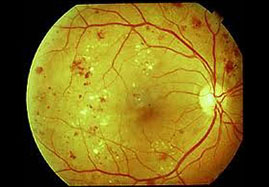
Diabetic Retinopathy
Approximately 6% of South Africans suffer with diabetes
Read More
With the highest prevalence being among Indians due to a strong genetic predisposition for diabetes. Diabetes mellitus may cause a reversible, temporary blurring of the vision, or it can cause a severe, permanent loss of vision. Diabetic retinopathy (damage to the small blood vessels in the retina) is a major cause of visual loss and a leading cause of blindness. Risk factors for diabetic retinopathy include the duration of diabetes, poor blood sugar control, hypertension, renal disease, high cholesterol and diabetes in pregnancy. All diabetic patients must have a dilated eye examination at least once a year in order to detect and timeously treat complications of diabetes in the eye. Treatment options include laser, intravitreal eye injections of anti-VEGF agents, and surgery for advanced diabetic retinopathy. The ideal treatment is primary prevention with lifestyle modifications such as a healthy diet and exercise; blood glucose, blood pressure and cholesterol control in diabetic patients as well as annual screening for diabetic retinopathy.
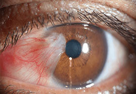
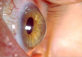
Pterygium
Pterygium is a growth on the cornea and the conjunctiva
aaaa
Read More
These growths are believed to be caused by dry eye, exposure to wind and dust and UV or sun exposure. In many cases no treatment is needed. Sometimes eyedrops and ointments may be used to reduce inflammation. If the growth threatens sight or causes persistent discomfort, it can be surgically removed. The goal of pterygium excision is to decrease irritation/inflammation, achieve a normal, smooth ocular surface, improve the decreased vision caused by the pterygium, and prevent regrowth, if possible by using conjunctival transplantation. A conjunctival autograft involves moving a piece of your own conjunctiva (filmy white part of the eye) to the area where the pterygium is excised (removed). This technique may be used for the management of both primary and recurrent pterygium.
Keratoconus and corneal cross linking
Keratoconus is a progressive thinning of the cornea which becomes cone shaped
Read More
Although current treatments for keratoconus, such as contact lenses and corneal ring implants, can improve vision, they do not treat the underlying cause, corneal weakness. As such they do not prevent the keratoconus from progressing.
Corneal collagen cross-linking aims to address this problem. Corneal collagen cross-linking
(CXL) uses chemicals to form connections, or cross-links, between adjoining strands of collagen, which is an important part of the structure of the cornea. This is currently performed using riboflavin (vitamin B2) and ultraviolet light (UVA).It is important to understand that collagen cross-linking treatment is not a cure for keratoconus in that significant corneal distortion is expected to remain even after the treatment. Rather, it aims to slow or even halt the progression of the condition. After the treatment, it is expected that it will continue to be necessary for individual people to wear spectacles or contact lenses (although a change in the prescription, probably to a lower powered script, may be required). As such it is felt likely that the treatment will prevent further deterioration in vision and the need for corneal transplantation.
Other eye conditions
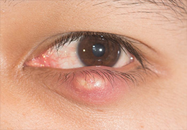
Chalazion
A chalazion is also known as a meibomian cyst
Red eye
The acute red eye is a frequent reason for patients seeking medical treatment
Read More
Common causes include allergic or infectious conjunctivitis, keratitis (corneal infections) episcleritis, uveitis and spontaneous or post traumatic subconjunctival haemorrhage
Chalazion
A chalazion, also known as a meibomian cyst, is a localized inflammatory response involving sebaceous glands of the eyelid that occurs when the gland duct is obstructed.
A chalazion may resolve spontaneously or with warm compresses, lid scrubs, and lid massage. When there is no improvement, the chalazion may be incised and drained. After local anaesthesia, a chalazion instrument is put in place and an incision is made in the inner aspect of the eyelid. The contents of the chalazion are then carefully drained with a curette followed by gentle pressure or heat to control any bleeding.
Patient Information Forms
Cataract surgery
Pterygium surgery
Corneal cross linking
Chalazion procedure
Your Health Starts Here
Flexible appointments and urgent care
Or call — 031 581 2546